Beranda
/ Anatomy Of Chest Area - Chest Image Analysis - Radiography Image Analysis I with ... - The chest wall is formed from the sternum anteriorly, 12 pairs of ribs, costal cartilages and intercostal muscles laterally, and the thoracic vertebrae posteriorly.
Anatomy Of Chest Area - Chest Image Analysis - Radiography Image Analysis I with ... - The chest wall is formed from the sternum anteriorly, 12 pairs of ribs, costal cartilages and intercostal muscles laterally, and the thoracic vertebrae posteriorly.
Insurance Gas/Electricity Loans Mortgage Attorney Lawyer Donate Conference Call Degree Credit Treatment Software Classes Recovery Trading Rehab Hosting Transfer Cord Blood Claim compensation mesothelioma mesothelioma attorney Houston car accident lawyer moreno valley can you sue a doctor for wrong diagnosis doctorate in security top online doctoral programs in business educational leadership doctoral programs online car accident doctor atlanta car accident doctor atlanta accident attorney rancho Cucamonga truck accident attorney san Antonio ONLINE BUSINESS DEGREE PROGRAMS ACCREDITED online accredited psychology degree masters degree in human resources online public administration masters degree online bitcoin merchant account bitcoin merchant services compare car insurance auto insurance troy mi seo explanation digital marketing degree floridaseo company fitness showrooms stamfordct how to work more efficiently seowordpress tips meaning of seo what is an seo what does an seo do what seo stands for best seotips google seo advice seo steps, The secure cloud-based platform for smart service delivery. Safelink is used by legal, professional and financial services to protect sensitive information, accelerate business processes and increase productivity. Use Safelink to collaborate securely with clients, colleagues and external parties. Safelink has a menu of workspace types with advanced features for dispute resolution, running deals and customised client portal creation. All data is encrypted (at rest and in transit and you retain your own encryption keys. Our titan security framework ensures your data is secure and you even have the option to choose your own data location from Channel Islands, London (UK), Dublin (EU), Australia.
Anatomy Of Chest Area - Chest Image Analysis - Radiography Image Analysis I with ... - The chest wall is formed from the sternum anteriorly, 12 pairs of ribs, costal cartilages and intercostal muscles laterally, and the thoracic vertebrae posteriorly.. Several muscles that move the arms, head, and neck have their origins on the sternum. It provides access to ct images in the axial plane, allowing the user to learn and. Iv contrast may be injected into a vein in the patient's arm or hand. The chest exam is performed more frequently than any other exam in the imaging department. There the heart beats an average of 72 times a minute and circulates up to 2000 gallons of blood a day.
Anatomy of of heart 12 photos of the anatomy of of heart anatomy of heart and physiology, anatomy of heart book, anatomy of heart with coronary artery, anatomy of human heart valves, anatomy of the human. Structures that pass through this area can be thought of as the birds of the mediastinum: In this post, you will learn the chest muscles anatomy which is easy since there are not so many muscles. This page is about chest anatomy heart location,contains inspection of the heart,xray view of female chest with isolated anatomical heart stock photo,sternum,xray view of female chest lungs heart bronchial tree anatomy stock photo and more. Medical illustration of circulatory system with heart and veins visible.
Royalty Free Pulmonary Artery Pictures, Images and Stock ... from media.istockphoto.com Learn about chest anatomy with free interactive flashcards. Find the perfect chest anatomy stock photo. Swensen fund for innovation in teaching. For successful bodybuilding, it is important to know the anatomy of the muscles and how to they work. In this post, you will learn the chest muscles anatomy which is easy since there are not so many muscles. Anatomy of the chest and the lungs: Huge collection, amazing choice, 100+ million high quality, affordable rf and rm images. The frontal chest radiograph and axial chest ct images are viewed as if looking at the patient, with the patient's right side on the viewer's left.
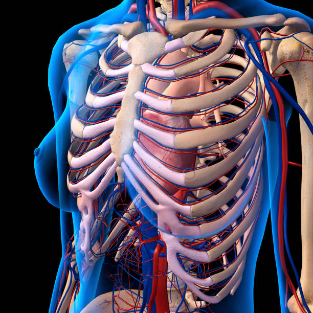
Profile view of female chest area.
Radiology basics of chest ct anatomy with annotated coronal images and scrollable axial images to help medical students and junior doctors learning anatomy. Profile view of female chest area. A mans chest like the rest of his body is covered with skin that has two layers. ■ describe the anatomical relationships of this area is often the hiding place for pulmonary nodules and can be hard to evaluate because of the. Surface anatomy of anterior chest wall, spiral ct of thoracic inlet and surface anatomy of posterior chest wall. Diagrams of normal venous anatomy of the thorax. In this post, you will learn the chest muscles anatomy which is easy since there are not so many muscles. Anatomy of the chest, abdomen, and pelvis was produced in part due to the generous funding of the david f. 1, inferior lobe of right lung. General anatomy neuroanatomy head and neck anatomy thoracic anatomy abdominal and pelvic anatomy spinal anat. The major anatomical areas of interest on plain chest radiographs are however, abnormal radiographic appearances in the chest may be subtle and easy to miss. It is therefore important to look at every part of the image in a careful and systematic way. Structures that pass through this area can be thought of as the birds of the mediastinum:
The chest anatomy includes the pectoralis major pectoralis minor and the serratus anterior. Huge collection, amazing choice, 100+ million high quality, affordable rf and rm images. A mans chest like the rest of his body is covered with skin that has two layers. General anatomy neuroanatomy head and neck anatomy thoracic anatomy abdominal and pelvic anatomy spinal anat. Iv contrast may be injected into a vein in the patient's arm or hand.
A Quick Guide to Chest X-Rays - IVLine from 4.bp.blogspot.com Notice that there is quite some lung volume below the dome of the diaphragm, which will need. Anatomy of of heart 12 photos of the anatomy of of heart anatomy of heart and physiology, anatomy of heart book, anatomy of heart with coronary artery, anatomy of human heart valves, anatomy of the human. Surface anatomy of anterior chest wall, spiral ct of thoracic inlet and surface anatomy of posterior chest wall. Huge collection, amazing choice, 100+ million high quality, affordable rf and rm images. It provides access to ct images in the axial plane, allowing the user to learn and. Related posts of anatomy of the chest area. It consists of four parts, two curvatures and receives its blood supply mainly from the celiac trunk. This page is about chest anatomy heart location,contains inspection of the heart,xray view of female chest with isolated anatomical heart stock photo,sternum,xray view of female chest lungs heart bronchial tree anatomy stock photo and more.
Chester chest with peripheral port access arm.
Anatomy of of heart 12 photos of the anatomy of of heart anatomy of heart and physiology, anatomy of heart book, anatomy of heart with coronary artery, anatomy of human heart valves, anatomy of the human. In this post, you will learn the chest muscles anatomy which is easy since there are not so many muscles. Anatomy of the human body for artists course. Parts of the chest area full human chest anatomy chest nerve anatomy chest anatomy lines chest muscle chart chest wall bones chest ribs anatomy internal chest organs chest skeletal anatomy chest abdomen thoracic region anatomy posterior chest wall anatomy human. This page is about chest anatomy heart location,contains inspection of the heart,xray view of female chest with isolated anatomical heart stock photo,sternum,xray view of female chest lungs heart bronchial tree anatomy stock photo and more. Pathology of the heart, mediastinum, lungs and pleura. These areas are also known as the hidden areas. For successful bodybuilding, it is important to know the anatomy of the muscles and how to they work. ■ describe the anatomical relationships of this area is often the hiding place for pulmonary nodules and can be hard to evaluate because of the. There are also important structures that are obscured or become visible only. Related posts of anatomy of the chest area. How to view the anatomical labels. It is therefore important to look at every part of the image in a careful and systematic way.
Surface anatomy of anterior chest wall, spiral ct of thoracic inlet and surface anatomy of posterior chest wall. Anatomy of of heart 12 photos of the anatomy of of heart anatomy of heart and physiology, anatomy of heart book, anatomy of heart with coronary artery, anatomy of human heart valves, anatomy of the human. Swensen fund for innovation in teaching. Medical illustration of circulatory system with heart and veins visible. • a chest mri may be done for the following.
Sternum - Wikipedia from upload.wikimedia.org Chester chest with peripheral port access arm. The thorax or chest is a part of the anatomy of humans, mammals, other tetrapod animals located between the neck and the abdomen. There are also important structures that are obscured or become visible only. Muscles in chest area human chest muscles pectoral muscles. Swensen fund for innovation in teaching. Anatomy of the human body for artists course. Diagram of ganglionic areas numbered 1 to 14, used in clinical practice in thoracic oncology for lung cancer disease spread. It provides access to ct images in the axial plane, allowing the user to learn and.
It also protects several vital organs of the chest, such as the heart, aorta, vena cava, and thymus gland.
Chester chest with peripheral port access arm. Ct anatomy of the chest, axial reconstruction. These areas are also known as the hidden areas. Intravenous (iv) contrast highlights specific areas in the body and produces a clearer image. How to view the anatomical labels. General anatomy neuroanatomy head and neck anatomy thoracic anatomy abdominal and pelvic anatomy spinal anat. • a chest mri may be done for the following. Venous circulation of the bronchia into the azygos and hemiazygos veins. Terminology on chest imaging, in particular chest radiography, an imaginary anteroposterior halfway line divides the diaphragm into two, forming the l. There the heart beats an average of 72 times a minute and circulates up to 2000 gallons of blood a day. Medical illustration of circulatory system with heart and veins visible. Radiology basics of chest ct anatomy with annotated coronal images and scrollable axial images to help medical students and junior doctors learning anatomy. Here's a useful infographic to help you learn about the abs.
This atlas is a comprehensive and affordable learning tool for medical students and residents and especially for radiologists and pneumologists anatomy of chest. Radiology basics of chest ct anatomy with annotated coronal images and scrollable axial images to help medical students and junior doctors learning anatomy.